


Arthrosis of the joints is a chronic disease characterized by the development of degenerative changes in the articular cartilage, as a result of which the bone tissue is deformed. The joints of the big toes, hip and knee joints are most often affected.
Symptoms of the disease
- The first clinical symptom of arthrosis is pain in the affected joint during excessive physical exertion. Painful sensations can occur during movement. As the disease progresses, joint pains bother a person even at rest and cause insomnia.
- Crunching joints. Due to the destruction of the cartilaginous layer, friction of the bones occurs, and when moving in the joint, clicks and crunch are heard. As the disease progresses, the crunch increases.
- Decreased mobility. If the joint is damaged, movements in it are limited, with severe arthrosis, the patient has stiffness of the limbs in the morning.
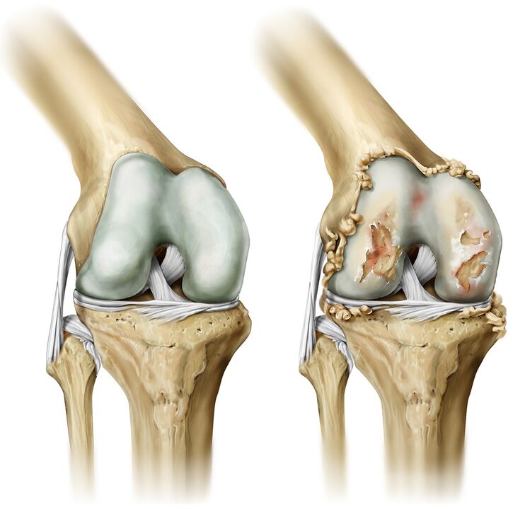
- Joint deformity. In the absence of adequate and timely treatment, the joint is deformed, its appearance changes.
- With an exacerbation of the inflammatory process, the patient has a decrease in the sensitivity of the toes and numbness of the fingertips.
Causes of the disease
The main reason for the development of arthrosis is the growth of the cartilage layer between the joint and the bone. The contributing factors are:
- Intense physical activity;
- Joint microtrauma;
- Frequent fractures
- Wearing tight shoes or high heels
- Congenital predisposition.
Diagnostics
The main method for diagnosing arthrosis is a carefully collected patient history (professional history).
The diagnosis is made on the basis of examination of the patient and additional studies, including X-ray of the joints, arthroscopy, ultrasound, MRI and computed tomography.
- Ultrasound. This research method is reliable and harmless. Since ultrasound diagnostics refers to non-invasive methods, this study has no contraindications. With the help of ultrasound, it is possible to diagnose thinning of cartilage tissue, degenerative changes in the articular menisci, thickening of the membranes of the joint, the presence of fluid in the joint cavity. This study allows you to accurately select a method for treating arthrosis.
- MRI and computed tomography. With the help of computed tomography and MRI, it is possible to assess the condition of the joint: the thickness of the cartilage, the presence of erosions or cysts in the bone tissue, to determine the amount of intra-articular fluid.
- Arthroscopy. This study is more often carried out to determine the cause of the development of arthrosis.
Complications
In the absence of timely medical care, arthrosis progresses and threatens with complications such as:
- Inflammation of the tissues around the joint;
- Limitation of mobility of the affected joints;
- Degenerative changes in the hip joint;
- Changing the shape of the joints.
Treatment of the disease
Treatment is prescribed to the patient depending on the degree of joint damage. Therapy for arthrosis begins with the relief of pain.
In parallel with analgesics, the patient is prescribed anti-inflammatory drugs. In addition to drug treatment, the patient undergoes a course of physiotherapy.
Massage of the affected limbs after the acute form of the inflammatory process has subsided can reduce pain, normalize joint mobility, and relieve muscle spasm.
Physiotherapy exercises are prescribed to relieve stiffness in the muscles, warm them up and strengthen the general condition of the patient. Exercise helps to maintain correct posture and an even gait.
Sanatorium treatment is indicated in the period of stable remission. Mud baths, applications and other procedures help to restore the motor function of the joints and relieve pain.
If conservative methods of treatment do not bring the expected effect, then the patient is prescribed surgical joint replacement. Endoprostheses are made from a material that is not rejected by the human body. They allow you to fully restore the physiological functions of the affected joint.
Unique treatments: radiofrequency ablation and disruption of the integrity of the method by disrupting the integrity of the nerve that causes pain.
Risk group
The risk group includes people:
- Overweight;
- Varicose veins;
- Athletes;
- Pianists;
- Programmers.
Prophylaxis
Prevention of arthrosis is as follows:
- Good nutrition;
- Prevention of injuries and fractures;
- Limiting the load on the joints with a hereditary predisposition;
- Body weight control;
- Wearing shoes that fit.
Diet and lifestyle
With a hereditary predisposition to the development of arthrosis, as well as during an exacerbation of the disease, it is necessary to adjust the diet. It is recommended to include in the diet sea fish (sardines, salmon, tuna), fresh vegetables and fruits, cereals. Limit baking, fatty meats, chocolate, and alcohol.
It is recommended to spend more time in the fresh air and not to expose the joints to increased physical activity.